Thyroid gland calcitonin thyroglobulin follicles cells hormone histology antibody produced saccular healthjade The thyroid gland Histology slides database: histological diagrams of thyroid gland
Calcitonin function, where is calcitonin produced & calcitonin uses
The thyroid gland 17.4 the thyroid gland, the endocrine system, by openstax Calcitonin function, where is calcitonin produced & calcitonin uses
Thyroid gland calcitonin thyroglobulin hormone histology follicles location antibody saccular healthjade footnote
The thyroid glandThyroid gland cartilage part trachea body diagram around anterior tissue neck isthmus larynx figure located anatomy labeled posterior parathyroid physiology Parathyroid glands gland posterior thyroid micrograph small figure lm cells anatomy diagram tissue chief embedded blood surface physiology vessel partThyroid gland histology.
Thyroid gland anterior located jobilize cartilage superiorThyroid histology gland Thyroid gland location, function, hormones, problems and surgeryThyroid gland location, function, hormones, problems and surgery.

Thyroid gland labeled diagram histology cells tissues human kenhub
Pathology outlinesThyroid gland endocrine parathyroid labeled histology system 400x cells follicles la lab viewof amo here dysfunction tiroides ap school med Thyroid gland – veterinary histologyThyroid gland follicular epithelium follicles histology epithelial cuboidal ohiostate pressbooks cytoplasm lined single.
Thyroid gland: cells, tissues, labeled diagram (preview)Thyroid gland anatomy follicles function cells histology gross anterior calcitonin hormones hormone source location saccular showing note figure The endocrine system: thyroid and parathyroid glandGland thyroid histological histology diagrams slides.

Thyroid gland diagram labeled posterior isthmus tissue trachea cartilage left near parathyroid glands anterior its figure part located around under
Thyroid gland physiology neck trachea hormonesThe parathyroid glands Thyroid histology gland microscope slide slides endocrine labels cells tissue label tissues anatomy structures labeled normal system human pancreas histoThyroid gland histology..
Thyroid histology pathology outlines pigment brown perinuclear pathologyoutlines .


Calcitonin function, where is calcitonin produced & calcitonin uses

Thyroid gland – Veterinary Histology
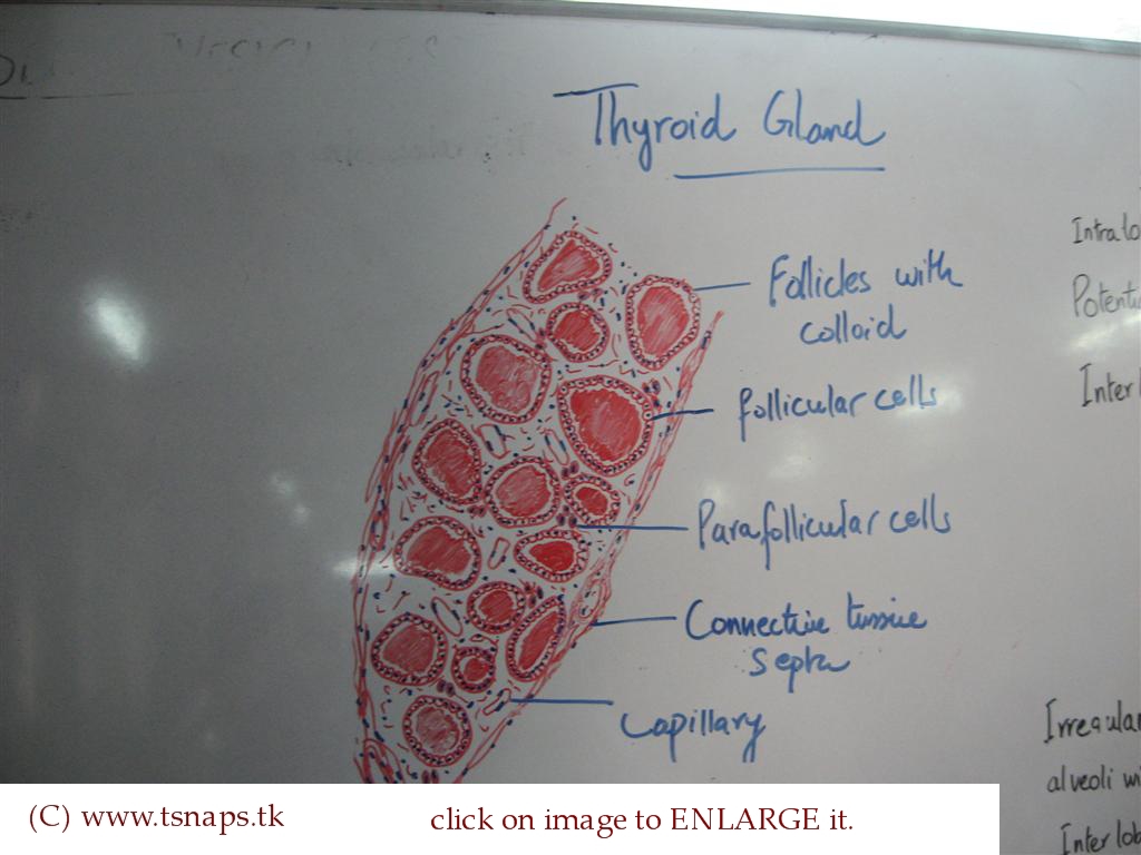
Histology Slides Database: histological diagrams of thyroid gland

The Endocrine System: Thyroid and Parathyroid Gland | Baseline of

The Thyroid Gland | Anatomy and Physiology

thyroid gland histology.

The Thyroid Gland | Biology of Aging

Thyroid Gland Location, Function, Hormones, Problems and Surgery

Pathology Outlines - Histology

